Leg Bone Diagram Labeled / Femur Bone High Res Stock Images Shutterstock / Diagram and names of leg bones, diagram of foot and leg bones, diagram of leg bones, diagram of lower leg bones, diagram of the bones in your leg, bone, diagram and.
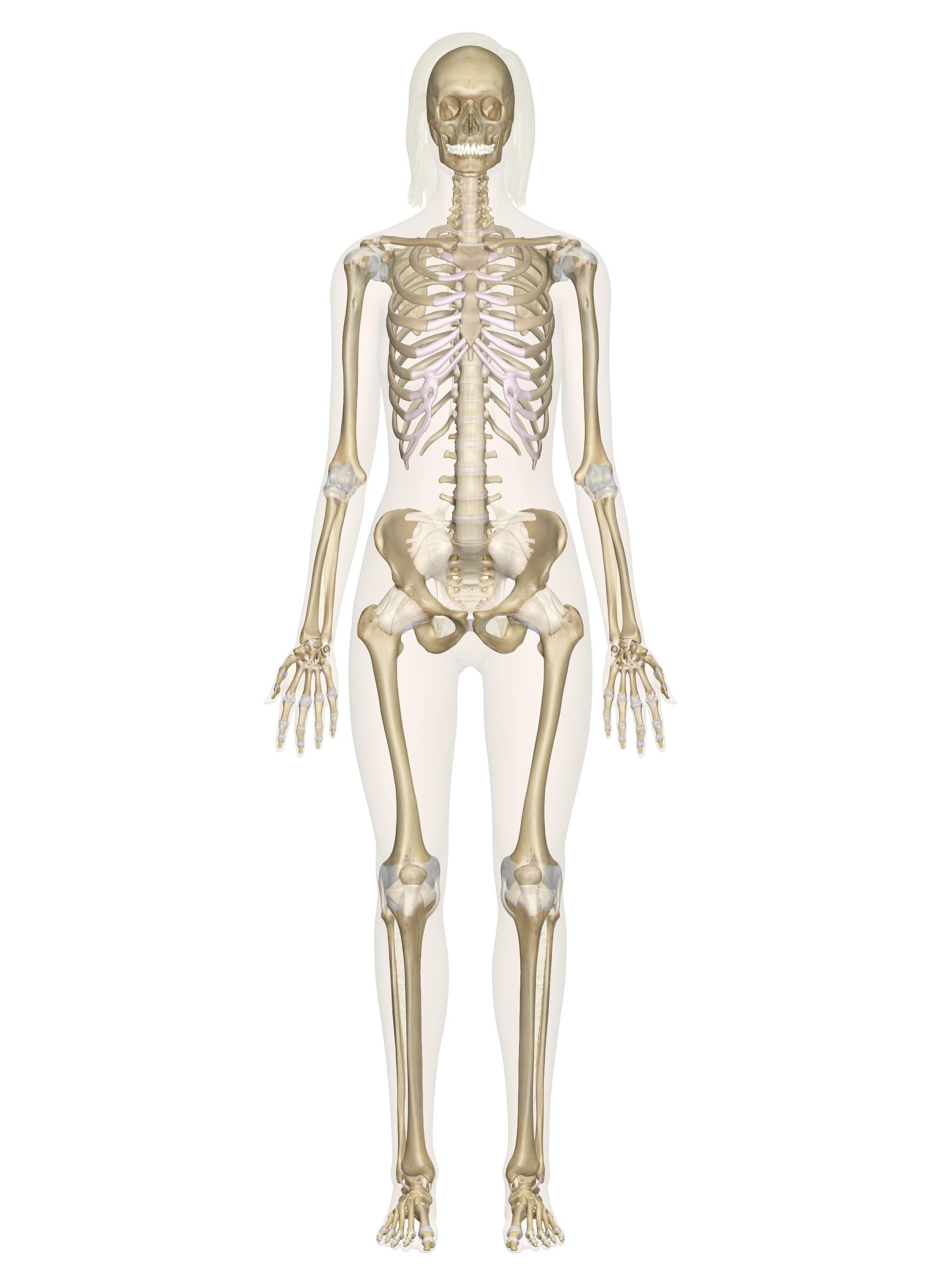
Leg Bone Diagram Labeled / Femur Bone High Res Stock Images Shutterstock / Diagram and names of leg bones, diagram of foot and leg bones, diagram of leg bones, diagram of lower leg bones, diagram of the bones in your leg, bone, diagram and.. The bony bumps (or protrusions) seen and felt on the ankle have their own names: Human skull anatomy bones of the skull with sections and internal views. The bones together make up the hip. The pubis, ischium, and ilium together constitute the pelvis while the thigh bone is the femur. This diagram of a feline skeleton shows you where all of your cat's bones are.
Labeled human leg bones created for use in leg bone. The bones of the hip include the femur, the ilium, the ischium, and the pubis. The bones together make up the hip. Human skull anatomy bones of the skull with sections and internal views. The foot bones shown in this diagram are the talus, navicular, cuneiform, cuboid, metatarsals and calcaneus.
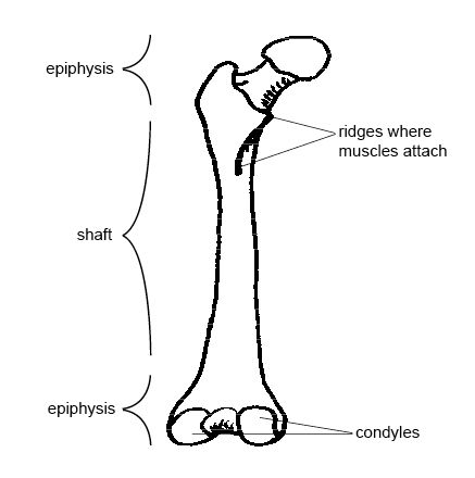
Knee Joint Picture Image On Medicinenet Com from images.medicinenet.com The bones of the leg and foot form part of the appendicular skeleton that supports the many muscles of the lower limbs. Knee, leg, and foot (overview) how many times have a layman's language and anatomy ever matched? The ankle is a large joint made up of three bones: The upper leg is often called the thigh. Dog leg anatomy just like humans have arms and legs dogs have forelegs and hind legs. Learn anatomy physiology leg bones with free interactive flashcards. Also called the thigh bone, this is the longest bone in the body.it. The thigh bone, or femur, is the large upper leg bone that connects the lower leg bones (knee joint) to the pelvic bone (hip joint).
It's the area that runs from the hip to the knee in each leg.
These muscles work together to produce movements such as standing, walking, running, and jumping. The bone at the top of the leg. Labeled human leg bones created for use in leg bone. In humans the neck of the femur connects the shaft and head at a 125 degree angle, which is efficient for walking. The human knee actually comprises of two joints, the Leg bone anatomy diagram diagram of human leg human anatomy human leg bones anatomy stock photo download image now anatomy of the knee central coast orthopedic medical group The pubis, ischium, and ilium together constitute the pelvis while the thigh bone is the femur. A leg bone is a bone found in the leg. Formed by the left and right hip bones, the pelvic girdle connects the lower limb (leg) bones to the axial skeleton. Diagram and names of leg bones, diagram of foot and leg bones, diagram of leg bones, diagram of lower leg bones, diagram of the bones in your leg, bone, diagram and. The bones of the leg are the femur, tibia, fibula and patella.the foot bones shown in this diagram are the talus, navicular, cuneiform, cuboid, metatarsals and calcaneus. At the same time, the bones and joints of the leg and foot must be strong enough to support the body. Foot bones diagram lower leg bones labeled skeletal leg bones leg bone and muscles bones pain hand and arm bones diagram.
The outer shin bone the knee bones work together to support the body and transfer forces between the hip and foot, allowing the leg to move smoothly and efficiently. Choose from 500 different sets of anatomy physiology leg bones flashcards on quizlet. Review date 7/8/2020 updated by: Below given knee diagram will help you to understand. Human skull anatomy bones of the skull with sections and internal views.
Skeletal System Labeled Diagrams Of The Human Skeleton from innerbody.imgix.net The lower leg is comprised of two bones the tibia and the smaller fibula. These muscles work together to produce movements such as standing, walking, running, and jumping. The lower thigh is the part of the hind leg beneath. A leg bone is a bone found in the leg. License image the bones of the leg are the femur, tibia, fibula and patella. Benjamin ma, md, professor, chief, sports medicine and shoulder service, ucsf department of orthopaedic surgery, san francisco, ca. The foot bones shown in this diagram are the talus, navicular, cuneiform, cuboid, metatarsals and calcaneus. In humans the neck of the femur connects the shaft and head at a 125 degree angle, which is efficient for walking.
This image is an edited version of this image that was created by user:ladyofhats (mariana ruiz villarreal).
The thigh bone, or femur, is the large upper leg bone that connects the lower leg bones (knee joint) to the pelvic bone (hip joint). The hock is the oddly shaped joint that makes a sharp angle at the back of the dogs legs. Bone diagram forehead (frontal bone) nose bones (nasals) cheek bone (zygoma) upper jaw (maxilla) lower jaw (mandible) breast bone (sternum) upper arm bone (humerus) lower arm bone (ulna) thigh bone (femur) collar bone (clavicle) toe bones (phalanges) ankle bones (tarsals) kneecap (patella) shin bone The main shin bone fibula: The rear legs of the dog begin with the femur bone which extends to a pair of bones known as the tibia and the fibula. Benjamin ma, md, professor, chief, sports medicine and shoulder service, ucsf department of orthopaedic surgery, san francisco, ca. Choose from 500 different sets of anatomy physiology leg bones flashcards on quizlet. Human skull anatomy bones of the skull with sections and internal views. The outer shin bone the knee bones work together to support the body and transfer forces between the hip and foot, allowing the leg to move smoothly and efficiently. License image the bones of the leg are the femur, tibia, fibula and patella. A leg bone is a bone found in the leg. The lower extremity, commonly referred to as the leg, contains four bones (the femur, the patella, the tibia, and the fibula) and bends at the hip, the knee, and the ankle. Numbered one through five the bone that sits behind the big toe is no.
Below given knee diagram will help you to understand. Formed by the left and right hip bones, the pelvic girdle connects the lower limb (leg) bones to the axial skeleton. Beside that, we also come with more related ideas as follows free printable human anatomy coloring pages, lower leg muscle diagram blank and lower limb bones unlabeled. Its lower end helps create the knee joint. Also called the thigh bone, this is the longest bone in the body.it.
Anatomy And Physiology Of Animals The Skeleton Wikibooks Open Books For An Open World from upload.wikimedia.org The rear legs of the dog begin with the femur bone which extends to a pair of bones known as the tibia and the fibula. The ankle is a large joint made up of three bones: This diagram of a feline skeleton shows you where all of your cat's bones are. I am not an expert at anatomy. Bone diagram forehead (frontal bone) nose bones (nasals) cheek bone (zygoma) upper jaw (maxilla) lower jaw (mandible) breast bone (sternum) upper arm bone (humerus) lower arm bone (ulna) thigh bone (femur) collar bone (clavicle) toe bones (phalanges) ankle bones (tarsals) kneecap (patella) shin bone The four knee bones fit together to make two different knee joints: The blood supply to and/or from the navicular bone is disrupted. It's the area that runs from the hip to the knee in each leg.
Human skull anatomy bones of the skull with sections and internal views.
The bones together make up the hip. The ankle is a large joint made up of three bones: The femur is the largest bone in the body and the only bone of the thigh. The four knee bones fit together to make two different knee joints: Beside that, we also come with more related ideas as follows free printable human anatomy coloring pages, lower leg muscle diagram blank and lower limb bones unlabeled. Benjamin ma, md, professor, chief, sports medicine and shoulder service, ucsf department of orthopaedic surgery, san francisco, ca. Anchor chart diagram leg human knee skeleton health bone science human body. Each leg is made up of four bones. I am not an expert at anatomy. Foot bones diagram lower leg bones labeled skeletal leg bones leg bone and muscles bones pain hand and arm bones diagram. Its lower end helps create the knee joint. This diagram depicts diagram leg bones anatomy.human anatomy diagrams show internal organs, cells, systems, conditions, symptoms and sickness information and/or tips for healthy living. Human skull anatomy bones of the skull with sections and internal views.
The knee joint is the largest joint in the body and is primarily a hinge joint, although some sliding and rotation occur leg bone diagram. The ankle is a large joint made up of three bones:


Post a Comment
0 Comments